- ANESTHESIOLOGY
- CARDIOLOGY
- Dental
- Department
- DERMATOLOGY
- ENT
- GASTROENTEROLOGIST
- GENERAL MEDICINE
- GENERAL SURGERY
- GYNECOLOGY
- MAXILLOFACIAL SURGEON
- Medical Gastroenterology
- NEPHROLOGY
- NEURO PHYSICIAN
- NEURO SURGERY
- NEUROLOGY
- ORTHAPEDIC
- PATHOLOGY
- PEDIATRICIAN
- PHYSIOTHERAPY
- PLASTIC SURGEON
- PULMONOLOGY
- RADIOLOGY
- SURGICAL ONCOLOGY
RADIOLOGY
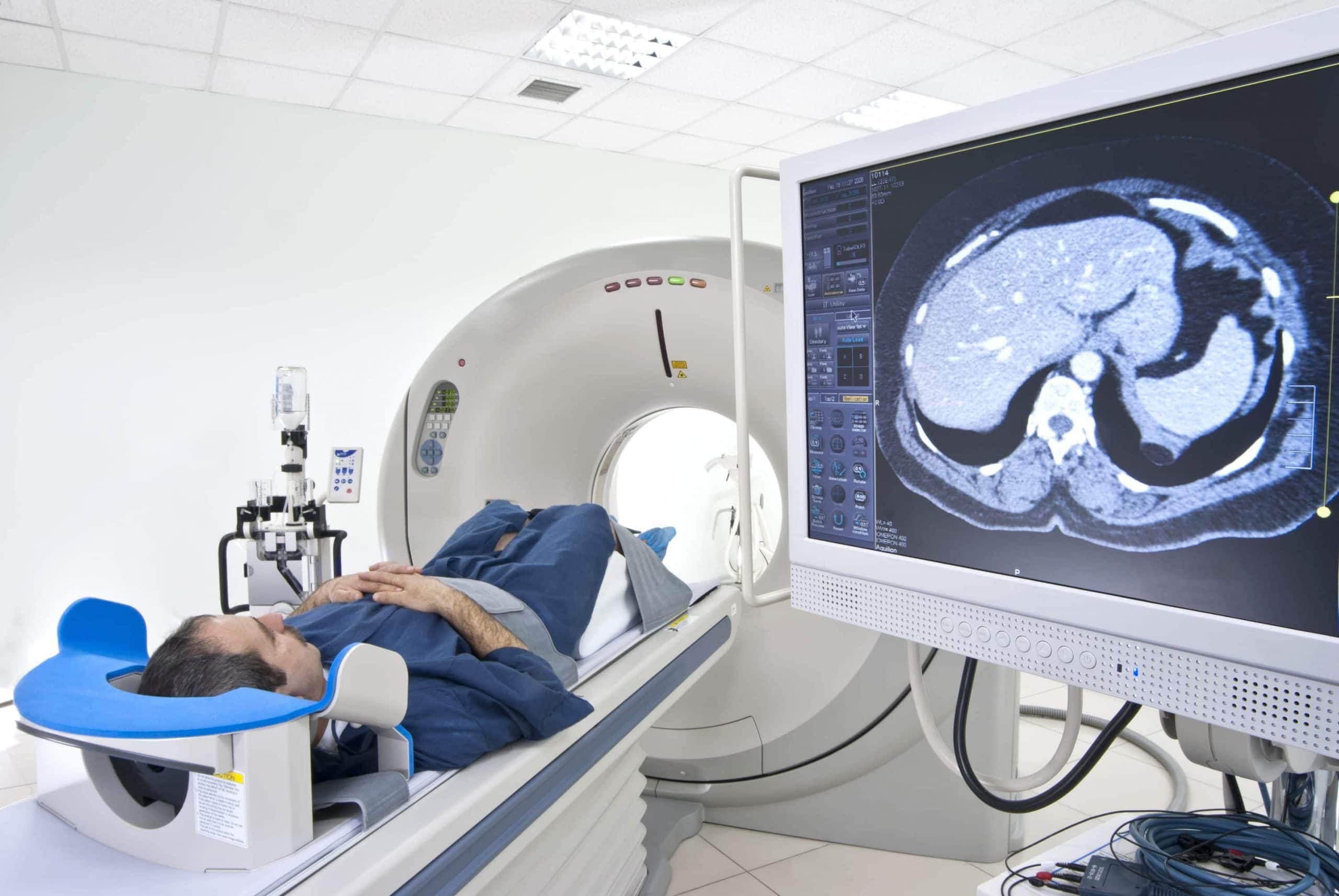
Radiology is a medical specialty that utilizes various imaging techniques to visualize internal structures, organs, and tissues within the human body. It plays a fundamental role in disease diagnosis, treatment planning, and monitoring across a wide range of medical disciplines, including radiography, computed tomography (CT), magnetic resonance imaging (MRI), ultrasound, nuclear medicine, and interventional radiology. In this comprehensive guide, we will delve into the fascinating world of radiology, exploring its history, principles, technologies, clinical applications, and future directions.
Historical Evolution
Early Beginnings
The origins of radiology can be traced back to the late 19th century with the discovery of X-rays by Wilhelm Conrad Roentgen in 1895. Roentgen’s accidental discovery of X-rays revolutionized medical imaging by enabling physicians to visualize internal structures and detect pathological changes without invasive procedures. The first X-ray image, a radiograph of Roentgen’s wife’s hand, revealed the bones and structures beneath the skin, sparking worldwide interest and enthusiasm for this groundbreaking technology.
Emergence of Radiography
Following Roentgen’s discovery, radiography quickly became a valuable diagnostic tool in medicine. Physicians and scientists around the world experimented with X-ray equipment, developing techniques for producing high-quality radiographic images of various body parts and anatomical structures. Radiography played a crucial role in diagnosing fractures, detecting foreign bodies, and localizing tumors, leading to significant advancements in orthopedics, surgery, and oncology.
Development of Computed Tomography (CT)
The introduction of computed tomography (CT) in the 1970s marked a significant milestone in radiology. CT imaging, invented by Godfrey Hounsfield and Allan Cormack, utilized computer algorithms to reconstruct cross-sectional images of the body from multiple X-ray projections, providing detailed anatomical information and improved tissue contrast compared to conventional radiography. CT scanning rapidly became an essential tool for diagnosing intracranial lesions, abdominal pathologies, and musculoskeletal disorders, revolutionizing diagnostic radiology and patient care.
Magnetic Resonance Imaging (MRI) Revolution
The development of magnetic resonance imaging (MRI) in the 1980s further expanded the capabilities of diagnostic radiology. MRI, based on the principles of nuclear magnetic resonance (NMR), utilizes strong magnetic fields and radiofrequency pulses to generate detailed images of soft tissues, organs, and functional brain anatomy. MRI offers superior soft tissue contrast, multiplanar imaging capabilities, and functional imaging modalities such as diffusion-weighted imaging (DWI), functional MRI (fMRI), and magnetic resonance spectroscopy (MRS), making it indispensable for diagnosing neurological disorders, musculoskeletal injuries, and oncological conditions.
Principles of Medical Imaging
Ionizing Radiation
Ionizing radiation, including X-rays and gamma rays, is used in diagnostic radiology to produce images of the human body. When X-rays pass through tissues, they are absorbed, scattered, or transmitted based on the density and composition of the tissues encountered. Radiographic images are created by detecting the transmitted X-rays using specialized detectors, such as film, digital sensors, or image intensifiers, and converting the transmitted radiation into visible images.
Contrast Agents
Contrast agents are substances administered to patients to enhance the visibility of specific tissues, organs, or pathological processes during medical imaging procedures. Contrast agents can be administered orally, intravenously, or intrathecally, depending on the imaging modality and anatomical region of interest. Common contrast agents used in radiology include iodinated contrast media for CT imaging, gadolinium-based contrast agents for MRI, and ultrasound contrast agents for echocardiography and vascular imaging.
Image Acquisition Techniques
Medical imaging modalities utilize different techniques for image acquisition, each offering unique advantages and applications in clinical practice. Radiography and fluoroscopy use X-ray beams to produce two-dimensional (2D) projection images of the body, providing information about bone density, soft tissue density, and anatomical relationships. CT imaging utilizes X-ray beams and computer algorithms to generate cross-sectional images (slices) of the body, allowing for detailed visualization of internal structures and three-dimensional (3D) reconstructions.